The Human Body
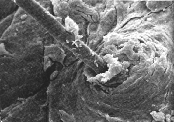 Figure 1: A close look at a single human hair. Just below the surface of the skin, within the hair follicle, are located sebaceous glands. They generate a fatty material call sebum, which oozes up the shaft to lubricate the hair and surrounding skin. A clump of extruded sebum can be seen around the base of the hair. 1000X-Dr. Joseph Gennaro, New York University (Gilmore, 1974).
 Figure 2: Inside the ear. The fan-like clusters at the top are made up of inner hair cells; the V's at the bottom are outer hair cells. They rest on a complicated structure called the organ of Corti deep within the inner ear. 7,800X-Dr. Goran Bredberg, Akademisda Sjukhuset, Uppsala, Sweden (Gilmore, 1974).
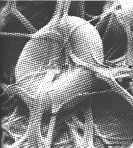 Figure 3: This striking picture shows the beginning of a vital process. The red blood cell is being enmeshed in a tangle of fibrin. This is the beginning of a blood clot, which occurs when the blood is exposed to air. 26,000X-Dr. Emil Bernstein, Eila Kairinen, Gillette Research Institute (Gilmore, 1974).
 Figure 4: This is a picture of a neuron from a sea snail. It shows the connection between the presynaptic membrane and the postsynaptic membrane of the cell body. 8000X-Dr. Edwin R. Lewis, Dr. Yehoshua Y. Zeevi, Dr. Thomas E. Everhart, University of California, Berkeley (Gilmore, 1974).
emooney@vt.edu 4/28/96
http://www.eng.vt.edu/eng/materials/classes/MSE2094_Notebook/
96ClassProj/experimental/human.html
Project Homepage | Experimental Page | Scanning Electron Microscope